
|
Home | MS.CHEST | Expert | Upload | My orders |
|
 中文
中文 EN
EN

|
Health Prophet
|
|
|
Health Prophet
|
|
|
Health Prophet
|
This Usage Agreement (hereinafter referred to as "this Agreement") specifies the conditions of use of the SATO radiology service (hereinafter referred to as "this Service") provided by MS.CHEST (Dalian) Co., Ltd. (hereinafter referred to as "this Company") on this website. Users who log in (hereinafter referred to as "Users") are required to use this Service in accordance with this Agreement.
Article 1 (Application)
1. This Agreement applies to all situations arising from the use of this Service by Users and this Company.
2. In addition to this Agreement, this Company has also separately formulated various agreements, such as usage rules (hereinafter referred to as "Individual Provisions"), regarding the provision of this Service. Regardless of their name, such Individual Provisions constitute part of this Agreement.
3. In the event of any ambiguity between this Agreement and the Individual Provisions referred to in the preceding paragraph, the Individual Provisions shall take precedence unless otherwise agreed.
Article 2 (Login)
1. Before using this Service, Users must first agree to the relevant provisions of this Agreement and then apply for login according to the methods specified by this Company. Once this Company confirms and agrees to the application, it is considered to be successful.
2. If any of the following circumstances arise when a User logs in, this Company may refuse the login application and shall not be responsible for informing the User of the reason for refusal.
1) False information is provided during the application process.
2) The applicant has a history of violating this Agreement.
3) Other circumstances that this Company deems inappropriate for the applicant to apply for login.
Article 3 (Management of Account Name and Password)
1. Users shall be responsible for managing the login account name and password provided by this Service.
2. Under no circumstances may Users transfer or lend their account name and password to third parties for use or sharing. When the account name and password used to log in to the system are consistent with the system login information, this Company considers that the User is using this Service.
3. This Company shall not be liable for any damages caused by the use of the account name and password by a third party, except in cases of intentional or gross negligence by this Company.
Article 4 (Service Contract)
1. This Service Contract is established once a User applies to this Company for X-ray consultation service in the manner specified by this Company, and this Company agrees to and notifies the applicant.
2. If this Company finds that a User falls under any of the following circumstances, this Company may terminate this Service Contract without prior notice.
1) The User violates the relevant provisions of this Agreement.
2) This Company is unable to provide consultation services due to problems with X-ray clarity or file format, etc.
3) Other circumstances that this Company considers to have broken the trust relationship between the Company and the User.
3. Matters related to the settlement method of this service, delivery method of X-rays, notification method of consultation results, cancellation method of service application, and refund method shall be implemented in accordance with the methods specified by the Company.
4. When the Company submits the reading results of the X-ray films provided by the user, it is deemed that the Company has fulfilled its obligations for this service. The Company shall not provide any inquiries, consultations, or additional information regarding the reading results of the X-ray films. If there are any unclear points about the reading results, please consult the attending physician or other treating physicians.
Article 5 (Usage Fees, etc.)
1. As the consideration for using this service, the user shall pay the service usage fee to the Company in accordance with the Company's designated payment method and price list.
2. In case of delayed payment of the service usage fee by the user, the user shall pay the Company a delay damages fee at an annual rate of 4% (calculated on a daily basis).
Article 6 (Intellectual Property Rights)
Except for the parts of the original elements belonging to the user, the Company and the legitimate rights holders of the elements provider shall own the copyright and other intellectual property rights of the elements required for providing this service. The user shall not copy, reproduce, modify or reuse them without permission.
Article 7 (Prohibited Actions)
When using this service, the user shall not engage in the following actions:
1. Actions that violate laws or public order and morals
2. Actions related to criminal activities
3. Actions that infringe on the copyright, trademark rights, and other intellectual property rights contained in this service
4. Actions that disrupt or hinder the functionality of the Company's servers or network systems
5. Actions that use the information obtained from this service for commercial purposes or for use in medical litigation
6. Actions that hinder or may hinder the Company's provision of this service
7. Actions that violate or attempt to violate the rules of connection, etc.
8. Actions that collect or store personal information of other users
9. Actions that impersonate other users
10. Actions that directly or indirectly provide benefits to organized crime groups regarding the implementation of this service
11. Other actions deemed inappropriate by the Company
Article 8 (Termination of Service Provided)
1. In the event of the following situations, the Company may terminate or suspend all or part of the Services without prior notice to the User:
1) When the computer system used to provide the Services undergoes repair, inspection or updating;
2) When the Company finds it difficult to provide the Services due to force majeure events such as earthquakes, lightning, fires, power outages, changes in policies or notifications from administrative agencies, or other natural disasters;
3) When the computer or communication line stops due to accidents;
4) When the Company deems that it is unable to provide the Services for any other reason.
2. The Company shall not be liable for any damages or losses suffered by the User or any third party resulting from the termination or suspension of the Services for any reason whatsoever.
Article 9 (Restrictions on Use and Deletion of Login Information)
1. In the event of the following situations, the Company may restrict the User's use of all or part of the Services or delete the User's login information without prior notice:
1) When the User violates any of the provisions of this Agreement;
2) When the Company confirms that the login information provided by the User is false;
3) When the settlement credit card used by the User for login is no longer valid;
4) When the User fails to pay any fees or debts;
5) When the Company notifies the User and the User fails to respond within a reasonable time;
6) When the User has not used the Services for a certain period of time after the last use;
7) When the Company deems that the User's use of the Services is inappropriate.
2. The Company shall not be liable for any damages suffered by the User as a result of the actions taken by the Company based on the above situations.
Article 10 (Withdrawal)
Users may withdraw from the Services in accordance with the withdrawal procedures established by the Company.
Article 11 (Disclaimer of Warranty and Limitation of Liability)
1. The Company does not guarantee that the Services are free of defects (including security, reliability, accuracy, completeness, effectiveness, suitability for specific purposes, security deficiencies, errors or defects, infringement, etc.) in fact or in law.
2. The Company shall not be liable for any damages suffered by the User as a result of using the Services. However, if the contract between the Company and the User regarding the provision of the Services (including this Agreement) violates the Civil Code or the Consumer Protection Act, the exemption clause shall not apply. Nevertheless, the Company shall not be liable for any damages caused by its negligence (excluding gross negligence) or infringement of the User's rights resulting from non-performance of obligations or infringement of rights caused by special circumstances (including those foreseen or foreseeable by the Company or the User).
3. The Company shall not be responsible for any transaction, contact, or dispute related to the Services between Users or between Users and any third party.
Article 12 (Changes to the Services, etc.)
The Company may change the content of the Services or suspend the provision of the Services without prior notice to Users, and the Company shall not be responsible for any losses incurred by Users as a result thereof.
Article 13 (Changes to the Terms of Use)
The Company may change the provisions of these Terms of Use without notice to Users as necessary. If a User starts using the Services after these Terms of Use have been changed, the User shall be deemed to have agreed to the changed Terms of Use.
Article 14 (Handling of Personal Information)
The Company shall handle personal information obtained in connection with the provision of the Services in accordance with the Company's Privacy Policy and applicable laws and regulations.
Article 15 (Notice or Contact)
Notices or contacts between Users and the Company shall be made in the manner specified by the Company. If a User does not change their registered contact information in the manner specified by the Company, the contact address currently registered shall be deemed valid, and any notices or contacts sent to that contact address shall be deemed to have been received by the User when they are sent.
Article 16 (Prohibition of Transfer of Rights and Obligations)
Users may not transfer their status under this Agreement or their rights and obligations under these Terms of Use to any third party or guarantee them for any third party without the prior written consent of the Company.
Article 17 (Governing Law and Jurisdiction)
1. This Agreement shall be governed by the laws of China.
2. Any disputes arising out of or in connection with the Services shall be under the exclusive jurisdiction of the court in the location of MS.CHEST (Dalian) Co., Ltd.
|
|
Health Prophet
|
This Privacy Policy (the "Policy") sets forth the policies and practices of Shenyang Lujiao Technology Co., Ltd. (hereinafter referred to as the "Company") with respect to the collection, use, and protection of users' personal information in connection with the services provided by the Company on this website (hereinafter referred to as the "Services").
The Company understands the importance of personal information to users and will do its utmost to ensure the security and reliability of users' personal information. The Company is committed to maintaining the trust of users in the Company and adhering to the principles of consistent responsibilities, clear purposes, choice and consent, minimum necessary, ensuring security, openness and transparency, etc., to protect users' personal information. At the same time, the Company promises that it will adopt corresponding security protection measures to protect users' personal information in accordance with the security standards established by the industry.
Please read and understand this Policy carefully before using the Services.
Last Updated: May 10, 2023
Article 1 (Personal Information)
"Personal Information" refers to the "personal information" as defined in the Personal Information Protection Law, which means information relating to a natural person that can identify a specific individual through such information as name, date of birth, address, telephone number, contact address, and other descriptions, as well as information that can identify a specific individual as an individual through data related to appearance, fingerprints, voiceprints, medical insurance card numbers, and the like (personal identification information).
"Sensitive Personal Information" refers to the "sensitive personal information" as defined in the Personal Information Protection Law, including information such as biometric data, specific identity, medical and health information, and the like.
Article 2 (Collection Method of Personal Information)
When a user registers for and uses our services, we may ask for personal information such as name, date of birth, address, telephone number, email address, bank account number, credit card number, and ID card number. In order to provide X-ray interpretation services, we collect sensitive personal information such as X-rays and medical history with the user's separate (individual) consent. In addition, we may collect transaction records containing user personal information and settlement-related information between the user and our partners (including information providers, advertisers, and ad broadcasters, collectively referred to as "partners").
Article 3 (Purpose of Collection and Storage Period of Personal Information)
1. The purposes for which we collect and use personal information are as follows:
① To provide and operate our services;
② To provide X-ray interpretation services by Japanese Consultant;
③ To respond to user inquiries (including identity verification);
④ To send emails introducing new features, updates, promotions, and other services offered by our company that the user has used;
⑤ To maintain necessary communication such as important notices;
⑥ To refuse to provide services to users who violate specific usage rules or use our services for specific improper purposes;
⑦ To allow users to view and change their login information and usage status;
⑧ To request payment for fee-based services;
⑨ Including purposes directly or reasonably related to the above purposes.
2. In order to facilitate the user's repeated use of our services and to allow the user to confirm the results of past interpretation services after registering as a member, we will retain personal information during the user's membership period. If the user cancels their membership, we will immediately delete the saved personal information. However, the storage period of personal information provided overseas shall be subject to the provisions of Article 5.
Article 4 (Provision of Personal Information to Third Parties)
1. We will not provide personal information to third parties without the user's prior consent, except as required by law.
2. Notwithstanding the preceding paragraph, the provision of information to the following parties shall not be considered as provision to a third party.
① When we entrust the processing of personal information in whole or in part within the scope necessary to achieve the purpose of use;
② When providing personal information as a prerequisite for complying with this policy due to business succession related to merger and other reasons.
Article 5 (Provision of Personal Information Overseas)
1. With the individual consent of the user, the Company will provide the necessary minimum information of the user, including the user's X-ray image, gender, age, and medical history to the Japanese corporation MS.CHEST and its delegated third party for the provision of X-ray interpretation services to the user. The user's name, address, phone number, email address, bank account, credit card number, and ID number will not be provided to overseas parties.
2. The Company will take necessary measures to ensure that users' personal information receives the same level of protection as that in the People's Republic of China when providing information overseas.
3. As stipulated in Article 1, the overseas recipients will keep the received user personal information for the minimum necessary period, which is usually within 45 days from the service provision date of each X-ray interpretation service.
Article 6 (Management and Protection of Personal Information)
1. The Company has implemented security measures that meet industry standards to protect the personal information provided by users, prevent unauthorized access, public disclosure, use, modification, damage, or loss of data. The Company will take all reasonable and feasible measures to protect users' personal information.
2. The Company will take all reasonable and feasible measures to ensure that irrelevant personal information is not collected. The Company will only retain users' personal information for the necessary period required to achieve the purpose of this policy (except when the retention period needs to be extended or legal permission is obtained).
Article 7 (User Rights)
1. Except for situations specified in laws, regulations, or relevant policies/guidelines, when the Company receives a request for disclosure of personal information from a user, it will promptly disclose such information to the user.
2. If a user finds that the personal information kept by the Company is incorrect, the user can request modification, addition, or deletion (hereinafter referred to as "modification, etc.") of such information from the Company in accordance with the procedures established by the Company.
3. When the Company receives such a request from the user and deems it necessary, it will immediately modify the personal information.
4. When the Company performs modification, etc. in accordance with the preceding paragraph or decides not to perform modification, etc., it will promptly notify the user.
5. When a user requests to stop using or delete personal information due to its use beyond the scope of the purpose or acquisition by improper means of personal information or for other reasons specified in laws and regulations (hereinafter referred to as "stop using, etc."), the Company will conduct necessary investigations immediately.
6. When it is deemed necessary based on the investigation results of the preceding paragraph, the Company should immediately stop using the personal information.
7. Regardless of the preceding two paragraphs, when there are significant costs for stop using, etc., or when it is difficult to stop using the personal information, the Company will take alternative measures to protect the user's rights if such measures are available.
8. To ensure security, user rights actions under this Article must be made in writing or other ways to verify the user's identity. The Company will require users to verify their identity before processing their requests.
9. Regarding reasonable requests from users, the Company generally does not charge a fee. However, for requests that are repeatedly made, exceed reasonable limits, or involve significant costs, the Company may charge a certain cost fee at its discretion. For requests that are groundlessly repetitive, require excessive technical means, pose risks to the legitimate rights and interests of others, or are highly impractical, the Company may refuse to comply.
Article 8 (Changes to the Privacy Policy)
1. The contents of this policy may be changed without notifying users to the extent that such changes do not violate laws and regulations.
2. Unless otherwise agreed by the Company, the revised privacy policy will take effect from the time it is posted on this website.
Article 9 (Personal Information of Minors)
1. The Company's website and services are mainly targeted at adults. Children should not create accounts with personal information as the subject without the consent of their parents or guardians.
2. In cases where children's personal information is collected with the consent of their parents, the Company will only use or disclose such information in accordance with the law, with the explicit consent of parents or guardians, or when necessary to protect children.
3. Although the definition of "children" varies under different local laws and customs, the Company considers anyone under the age of 14 to be a child.
4. If the Company discovers that it has collected personal information of a child without obtaining verifiable consent from the child's parents in advance, it will make efforts to delete such data as soon as possible.
Article 10 (Consultation Office)
For inquiries related to the disclosure, modification, cessation of use, and other matters concerning personal information and this policy, please contact the following office:
Address: "Address in China"
Company Name: Shenyang Antler Technology Co., Ltd.
Responsible Department: Customer Consultation Office
Email: info@antler-tech.com.cn
Contact Number: (024)8291 5371
|
|
Health Prophet
|
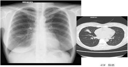
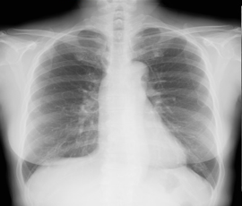
Interpretation of Chest X-Ray
There is a patchy shadow of approximately 3 cm in the lower right lung lobe.
The shadow is accompanied by a linear shadow on the outer lower edge.
Further examination using CT is necessary.
Interpretation of CT Chest X-ray:
A CT examination was subsequently performed.
The examination revealed a nodular shadow in the right middle lobe with a spiculated margin.
There was a strong suspicion of lung adenocarcinoma, so surgery was performed and the diagnosis was confirmed.
Because a lesion in the lung field (the black area) can be easily detected on a chest X-ray, this is a relatively easy case to detect.
However, it is difficult to distinguish between cancer and inflammatory conditions such as pneumonia based solely on a chest X-ray.
Original Japanese Text
胸部写真所見
右下肺野に約3㎝大の斑状影が見られます。
陰影の下外側に線状影を伴っています。
CTによる精査が必要です。
〈CT・胸部写真の解説〉
CTでは右中葉に結節影がみられ、結節周囲にスピクラを伴っています。
肺腺癌が強く疑われたため手術となり、肺腺癌であることが確認されました。
胸部写真にて肺野(黒い部分)に病変が認められるので、比較的発見は容易な症例です。
胸部写真のみでは、癌であるのか肺炎などの炎症であるのかの判断は困難です。
|
|
Health Prophet
|
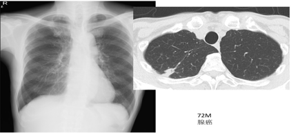
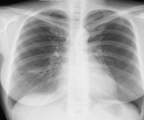
Interpretation of Chest X-ray
Reduced transparency is observed at the overlapping area of the first rib on the right side and the right clavicle.
Although it is possible to be post-tubercular inflammatory changes, further examination with CT is recommended.
〈Explanation of CT Chest X-ray〉
Nodular shadows are observed on the pleura of the right upper lobe according to the CT scan.
The CT scan can be tricky to distinguish between inflammatory changes and cancer.
Lung puncture was performed under CT guidance, and it was confirmed to be lung adenocarcinoma and underwent resection.
When the lesion overlaps with bones such as ribs, the heart, blood vessels such as the aorta and pulmonary artery, and the diaphragm, there is a higher likelihood of being overlooked.
Based on the chest X-ray, unlike Case 1, where the lesion was visible on both sides, in this case, only the difference between the left and right sides can be used to detect the lesion due to its overlap with bone.
Japanese Original
〈胸部写真所見〉
右第一肋骨と右鎖骨の重なる部位に透過性低下が見られます。
結核後などの炎症後変化の可能性はありますが、CTによる精査をお願いします。
〈CT・胸部写真の解説〉
CTにて右上葉胸膜に接して結節影が認められます。
CTでも古い炎症性変化と肺癌が悩ましい画像です。
CT下にて肺穿刺が行われ、肺腺癌であることが確認されたため、切除となりました。
肋骨などの骨、心臓、大動脈・肺動脈などの血管、横隔膜 などと病変が重なって見えている場合は、見落とされてしまう可能性が格段に上がります。
胸部写真にて骨に重なって病変が存在するので、症例1と異なり左右差のみが病変を見つけられるポイントになります。
|
|
Health Prophet
|
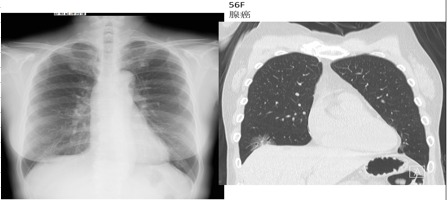
Interpretation of Chest X-ray
There is suspected blurring of the diaphragm line in the central part of the right diaphragm, and patchy shadows are observed in the upper part of the diaphragm.
For caution, a detailed and accurate CT examination is recommended.
<Interpretation of CT Chest X-ray>
The CT scan shows a circular ground-glass shadow above the diaphragm, and a nodular shadow can be seen in the central part.
This is a case that can be diagnosed as lung adenocarcinoma through CT.
This is a case that is difficult to detect through chest X-rays because the ground-glass shadow detected in the CT scan overlaps with the pulmonary artery, pulmonary vein, and other structures in the chest X-ray.
Only the "blurring" of the diaphragm line is the key to the discovery, making it a difficult case.
The key to discovering the case is the "blurred" diaphragmatic line, which is a difficult case.
Original Japanese Text
胸部写真所見
右横隔膜中央部に、横隔膜ラインの不明瞭化と、その上部に斑状影が疑われます。
念のためCTによる精査をお願いします。
〈CT・胸部写真の解説〉
CTにて横隔膜直上にすりガラス影が円形状に広がっており、中心部には結節影が見られます。CTでは肺腺癌と判断できる症例です。
胸部写真で発見するにはかなり困難な症例です。CTにて認められるすりガラス影は胸部写真では肺動脈・肺静脈などと多く重なっているためです。
横隔膜ラインの‘ボケ’のみが発見の決め手となる難しい症例でした。


 辽公网安备21010502000655号